Structural Organization in Animals: Class 11 Biology NCERT Chapter 7
Key Features of NCERT Solutions for Class 11 Biology Chapter 7 – Structural Organization in Animals
In the previous chapter: Anatomy of Flowering Plants, you will learn about Anatomy is the study of the Internal Structure of an organism. In this chapter of NCERT Class 11 Biology: Structural Organisation in Animals, you will study the world of animals and their internal and external features. After going through these notes made by our subject expert teachers, students will confidently identify the subject better and will be able to perform well in this subject.
Class 11 comes with new challenges that one may not be prepared for. In other words, it poses quite a number of difficulties in grasping the material. Our NCERT notes for Class 11 Biology Chapter 7 will help with it efficiently.
Quick revision notes
In multicellular living being a gathering of comparative cells alongside intercellular substances play out a particular capacity. Such association is called tissue.

Epithelial Tissue:
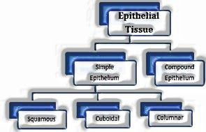
This tissue gives covering or coating to some piece of the body. Cells are minimally stuffed without intercellular space.
- The basic epithelium is made out of a single layer of cells and capacity as a coating of body depressions, tubes and ducts.
- The compound epithelium comprises of at least two than two layers of cells and has defensive capacity.
- The squamous epithelium is comprised of a single layer of levelled cells with unpredictable limits. They are available in the coating of veins, air sacs of lungs.
- Cuboidal epithelium is comprised of the single-layered 3D square like cells and found in channels of organs and rounded pieces of the nephron of kidney for ingestion and emission.
- Columnar epitheliums are comprised of tall and slim cells. The nuclei are situated at the base. The free surface may have microvilli found in the layer of the stomach and digestive tract. The ciliated one is called as ciliated epithelium.
- Columnar and cuboidal epithelium particular for discharge is known as the glandular epithelium, which might be unicellular as in cup cells of the nutritious trench or multicellular as in salivary organ
| Endocrine glands | Exocrine glands |
|
|
- The fundamental capacity of compound epithelium tissue is to give assurance against substance and mechanical pressure. They spread the dry surface of the skin, soggy surface of the buccal cavity, and so forth.
- Epithelial cells are held together by intercellular material to shape specific intersection.
Connective Tissues:
They are generally plentiful and broadly appropriated tissues which connection and bolster different tissues. Every single connective tissue with the exception of platelets, discharge strands of auxiliary protein called collagen or elastin to give versatility and adaptability.
- Loose Connective Tissues contain cells and filaments approximately orchestrated in semi-liquid ground substance. It incorporates areolar tissue and fat tissue.
| Areolar Connective Tissue | Adipose Connective Tissue |
|
|
- Dense connective tissue contains strands and fibroblast minimalistically pressed. The direction of strands might be an ordinary or sporadic example.
- In dense ordinary connective tissues, collagen filaments are available in columns between equal packs of strands as in ligaments and tendons.
| Tendon | Ligament |
|
|
- Cartilage, blood, and bones are special connective tissue.
| Cartilage | Bone |
|
|
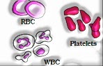
- Blood is liquid connective tissue containing plasma, red platelets, white platelets and platelets. It helps in the transportation of different substances between organs.
Muscle Tissue
- Each muscle is comprised of long round and hollow strands organized corresponding to one another. Filaments are made out of fine fibrils called myofibrils. Muscle filaments contract and unwind in light of incitement.
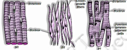
| Skeletal | Smooth | Cardiac |
|
|
|
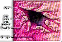
Neural Tissue
- The unit of the neural framework is the neuron. Neuroglial cell secures and underpins the neuron.
- At the point when neuron gets invigorated, electrical driving forces have produced that movement along with the plasma layer (axon).
The tissues sort out to shape organs which thus partner to frame organ framework in multicellular life forms.
Earthworm
- The earthworm is a ruddy earthy coloured earthbound invertebrate that lives in the upper layer of sodden soil. The regular Indian earthworms are Pheretima and Lumbricus.
- Worms have long round and hollow body separated into portions called metameres. The ventral surface contains genital pore and dorsal surface contain the mid-dorsal line.
- The first body section is called peristomium which contains the mouth. 14-16 fragments are secured by a dull band called the clitellum.
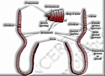
- The single genital pore is available on midventral line of fourteenth fragments. A couple of male genital pore is available on the eighteenth portion on ventro-horizontal side.
- All the fragment with the exception of first, last and clitellum contain S-molded setae for locomotion.
- The Alimentary canal is straight cylinder from first to last section having, buccal hole, solid pharynx, throat that prompts gizzards, which help in crushing the dirt particles and rotting leaves. Stomach and small digestive system prompts to the anus.
- Between 26-35 fragments, the digestive system has an inner middle crease called typhlosole. This builds the powerful zone of ingestion in the digestive system.
- The closed vascular framework comprises heart, veins and vessels. Blood organs are available on the fourth, fifth and sixth fragments. They produce blood cells and haemoglobin which is disintegrated in blood plasma.
- The earthworms need respiratory organs and breathe through clammy skin.
- Excretory organs are coiled segmental tubules known as nephridia. There are three sorts of nephridia: Septal nephridia, integumentary nephridia and pharyngeal nephridia.
- The sensory system is shown by ganglia ordered segment wise on the ventral matched nerve rope. The nerve rope in the front locale (third and fourth fragments) bifurcates and joins the cerebral ganglia dorsally to frame a nerve ring.
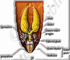
- The earthworm is hermaphrodite. Two sets of the testis are available in tenth and eleventh fragment. Prostrate and spermatic channel open to surface as a male genital pore on the eighteenth section.
- One set of ovaries is joined to the intersegmental septum of twelfth and thirteenth fragments. Female genital pore open on the ventral side of the fourteenth fragment. Shared trade of sperms happens during mating.
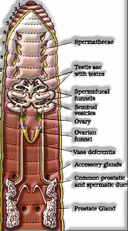
- Develop sperms and egg cells alongside nutritive materials are kept in a case in the dirt where treatment happens.
- Worms are known as companions of rancher since they make tunnels in the soil to make it permeable for breathing and root infiltration. Worms are likewise utilized for vermicomposting and as a snare in game fishing.
Cockroach(Periplaneta americana)
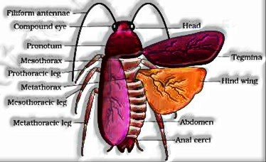
- Cockroaches are nighttime omnivorous life forms that live in sodden places all over the place. The assortment of cockroach is sectioned and distinct into head, chest and mid-region. The body is secured by the hard chitinous exoskeleton.
- Head is three-sided fit as a fiddle-shaped by combination of six sections to show adaptability. Head bears compound eyes. Receiving wire appended on head help in observing the earth.
- Chest comprises of three sections prothorax, mesothorax and metathorax. Forewings and rear wings are appended with the chest. Midsection comprises of 10 fragments.
| Male Cockroach | Female Cockroach |
|
|
Digestive System of Cockroach-
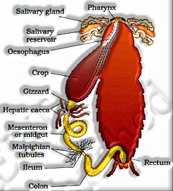
- The Alimentary channel is isolated into foregut, midgut and hindgut. Food is put away in the crop. Gizzard help in crushing the food particles.
- At the intersection of midgut and hindgut yellow-hued filamentous Malpighian tubules are available which help in discharge.
- Blood vascular framework is open sort having ineffectively evolved veins. The haemolymph is made of dreary plasma and haemocytes.
- The respiratory framework comprises of a system of the windpipe which opens through 10 sets of spiracles on the horizontal side.
- The sensory system of cockroach comprises of a progression of combined, segmentally orchestrated ganglia joined by matched longitudinal connectives on the ventral side. Three ganglia lie in the chest, and six in the mid-region. The sensory system of cockroach is spread all through the body.
- Each compound eye of cockroach comprises of around 2000 hexagonal ommatidia.
- With the assistance of a few ommatidia, a cockroach can get a few pictures of an article. This sort of vision is known as a mosaic vision with greater affectability yet less goal,
- Cockroaches are dioecious. The male regenerative framework comprises of a couple of testicles one lying on every parallel side in fourth sixth stomach portions. The female regenerative framework comprises of two huge ovaries arranged on second – sixth stomach sections.
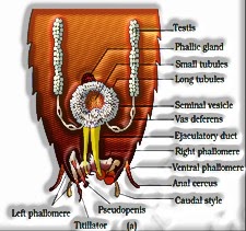
Male reproductive system / Female reproductive system
- The prepared eggs are encased in a container called oothecae. 9 to 10 oothecae are created by every female.
- Cockroaches are pests and destroy the food, contaminate with smelly excreta.
Frog (Rana tigrina)
Frogs are cold-blooded organism having the ability to change colours to hide from enemies. The body is divisible into head and trunk, bulged eyes covered by a nictitating membrane. Male frog is not the same as a female having vocal sacs and copulatory cushion on the first digit of the forelimb.
- Stomach related framework comprises of the nutritious channel and stomach related organs.
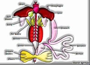
- Absorption begins in the stomach and last processing happens in the small digestive system. Processed food is consumed by villi and microvilli present in the inward mass of small digestive tract.
- Skin goes about as amphibian respiratory organs (cutaneous breath). On lands skin, buccal cavity and lungs go about as respiratory organs.
- The vascular arrangement of the frog is all around created closed sort. The heart is 3-chambered. Blood comprises of plasma, RBC, WBC and Platelets.
- Frogs have a lymphatic framework comprising of lymph, lymph channels and lymph hubs.
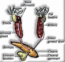
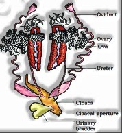
- The disposal of nitrogenous squanders is done by an all-around created excretory structure. The excretory framework comprises of a couple of kidneys, ureters, cloaca and urinary bladder. The frog discharges urea and hence is a ureotelic creature.
- The framework for control and coordination is exceptionally developed in the frog. It incorporates both the neural framework and endocrine organs
- Frogs have efficient male and female regenerative frameworks. Male conceptive organs comprise of a couple of yellowish ovoid testicles, which are found clung to the upper piece of kidneys by mesorchium.
- The female regenerative organs incorporate a couple of ovaries which are arranged close to kidneys.
- Fertilization is outer and happens in water. Advancement includes a larval stage called tadpole. Tadpole experiences transformation to shape the grown-up.
Reproductive systems of frog- Male / Female