Human Reproduction: Class 12 Biology NCERT Chapter 3
Key Features of NCERT Material for Class 12 Biology Chapter 3 – Human Reproduction
In the previous chapter, sexual reproduction in flowering plants you studied the different ways in which plants reproduce. In this chapter, Human reproduction, you will study that Propagation in people is by a sexual generation where both the male and female gametes treat to offer ascent to an incipient organism. The preparation of human incipient organism happens inside the body of the female. In this way, it is called Internal Fertilization.
Moreover, People are viviparous life forms who offer ascent to Embryos straightforwardly as opposed to laying eggs. Let us comprehend somewhat more about the male and female conceptive organs and the process of treatment in them.
Birth is actually the most excellent thing in this world. The way that we were all once a solitary cell and now as tall as a cabinet demonstrates how fascinating life seems to be. Human proliferation is a mysterious encounter. We get so cheerful when another part shows up in the family, isn’t that right? We play with him/her throughout the day. So the inquiry is: Do you need to find out about this otherworldly process inside and out? Truly! At that point plunge into the articles underneath.
Quick revision notes
People are explicitly recreating and viviparous. The regenerative occasions in people incorporate the development of gametes (gametogenesis), i.e., sperms in males and ovum in females, move of sperms into the female genital plot (insemination) and combination of male and female gametes (treatment) prompting arrangement of the zygote. This is trailed by arrangement and improvement of the blastocyst and its connection to the uterine divider (implantation), early-stage advancement (development) and conveyance of the child (parturition)
The Male Reproductive System
It cChapteronsists of:
- a) Primary sex organs for example a couple of testicles suspended in a scrotum.
- b) Secondary sex organs, for example, a couple of conduits each separated into rete testis, vasa efferentia, epididymis and vas deferens, ejaculatory channel and the related glands
- c) External genitalia
- The testicles are arranged outside the stomach cavity in a pocket called the scrotum, which helps in keeping up the low temperature of testicles vital for spermatogenesis.
- Although, Each testicle has around 250 testicular lobules and each lobule contain exceptionally curled seminiferous tubules in which sperms are delivered. Each seminiferous tubules is lined by two kinds of cells, spermatogonia ( male germ cell) and Sertoli cells.
- Leydig cells or interstitial cells present around the seminiferous tubules integrate and emit the androgen hormone.
 Ejaculatory channel store and transport the sperm from testicles to outside through urethra which starts from the urinary bladder and stretches out through the penis to its outer opening urethral meatus.
The penis is male outside genitalia. The extended finish of the penis is called the glans penis is secured by a free overlap of skin called a prepuce.
Male accessory glands incorporate combined fundamental vesicles, prostrate and matched bulbourethral glands. Emission of these glands shapes the fundamental plasma which contains fructose, calcium and proteins. The emission of bulbourethral glands additionally helps in the grease of the penis.
The Female Reproductive System:
It comprises of :
a)The essential sex organ that is a couple of ovaries
b)Secondary sex organs-the pipe framework comprising of a couple of fallopian tube, an uterus, cervix and vagina
c)External genitalia
d)Mammary glands
Ovaries are essential female sex organ that produces the female gamete and a few steroid hormones. Each ovary is secured by slight epithelium which encases the ovarian stroma, which is isolated into a fringe cortex and an inward medulla.
Fallopian tube reaches out from fringe of the ovary to the uterus. The part closer to the ovary is a channel moulded structure called infundibulum having finger-like projection called fimbriae.
Infundibulum prompts ampulla and gets together with uterus with the isthmus. The uterus is pear-formed structure likewise called belly.
Uterus open vagina through a restricted cervix. The cavity of the cervix (a cervical waterway) alongside vagina frames the birth channel.
The mass of uterus has three layers of tissue:
Perimetrium-outer film.
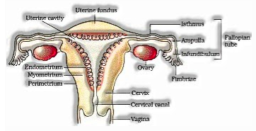
Myometrium – centre thick layer of smooth muscles which show solid compression during conveyance of child.
Endometrium – line the uterine divider and experience cyclic changes during the menstrual cycle.
Female outer genitalia incorporates
Mons pubis – pad of greasy tissues secured by the skin and pubic hair.
Labia majora-beefy overlay that encompasses the vaginal opening.
Labia Manora – matched overlay of tissue under labia majora.
The opening of the vagina is regularly in part secured by a layer called the hymen. The minuscule finger-like projection present at the upper intersection of two labia manora over the urethral opening is called the clitoris.
Mammary glands are matched structures that contain glandular tissues and variable fats. Each glandular tissue contains 15-20 mammary projections containing alveoli that discharge milk. Mammary conduits join to frame mammary ampulla.
Gametogenesis:
The process of development of male and female gametes in testicles and ovary individually is called gametogenesis. It is of two kinds:
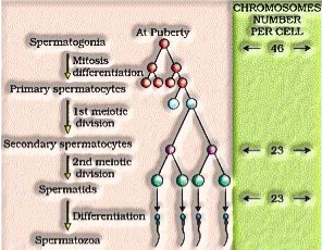
Spermatogenesis-in testicles youthful, male germ cells (spermatogonia) produce sperm by spermatogenesis that starts at adolescence.
The spermatogonia present at the internal side of seminiferous tubules duplicate by mitotic division and increment in number. Each spermatogonium contains 46 chromosomes.
Spermatogonia structures spermatocyte that experience meiotic division to imitate auxiliary spermatocytes having 23 chromosomes.
The spermatids are changed into spermatozoa by the process called spermiogenesis. The sperm heads stay inserted in Sertoli cells and are delivered from seminiferous tubules by the process of spermiation.
Hormonal control of spermatogenesis
Spermatogenesis started because of increment in the emission of gonadotropin delivering hormone by nerve centre
Increment in GnRH follow up on front pituitary and invigorate discharge of two gonadotropins, LH and FSH
LH follows up on Leydig cells and invigorates them to discharge androgens.
FSH follows up on Sertoli cells, invigorates discharge of certain variables which help in spermiogenesis
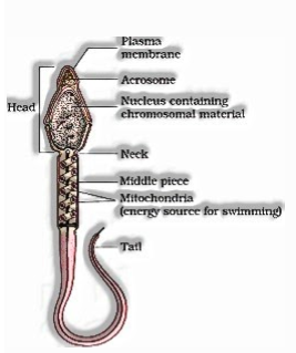
Structure of sperm
sperm is a minute structure made out of a head, neck, a centrepiece and a tail. Furthermore, The sperm head contains the lengthened haploid core, a front segment of which is secured by top like structure acrosome.
 Human male discharges around 200-300 million sperms during a Copulation. The original plasma alongside the sperms establishes the semen. The capacity of male sex auxiliary conduits and glands are kept up by androgen hormones.
Oogenesis: The process of arrangement of developing female gametes is called oogenesis. It began during the undeveloped improvement stage when a great many oogonia (gamete mother cells) are shaped in each fetal ovary.
The gametes mother cells start division and go into prophase-I of meiotic division and get transiently captured at that stage called essential oocytes.
Each essential oocyteget encompassed by a layer of granulosa cell than it is called the essential follicle.
At adolescence, around 60,000-80,000 essential follicles are left in each ovary
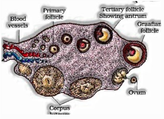
So, Essential follicle gets encompassed by more layers of granulosa cells called optional follicle that change into tertiary follicle that contains liquid-filled pit called the antrum.
The tertiary follicles further change into the develop follicle called Graafian follicle, which joy to deliver optional oocytes (ovum) from the ovary by the process of ovulation.
Menstrual cycle:
The conceptive cycles in female primates is called the menstrual cycle. It starts at adolescence and is called menarche.
Periods of Menstrual Cycle
The menstrual cycle comprises of the following four stages:
(1) Menstrual Phase:
(I) In a 28 days menstrual cycle, the menses happens on cycle days 3-5.
(ii) The creation of LH from the front projection of the pituitary organ is diminished.
(iii) The withdrawal of this hormone causes degeneration of the corpus luteum and, hence progesterone creation is diminished.
(iv) Production of estrogen is additionally diminished in this stage.
(v) The endometrium of uterus separates and the feminine cycle starts.
(vi) The cells of endometrium discharges, blood and unfertilised ovum comprise the menstrual stream.
(2) Follicular Phase:
(I) This stage normally incorporates cycle days 6-13 or 14 out of a 28 days cycle.
(ii) The follicle invigorating hormone (FSH) emitted by the foremost projection of the pituitary organ animates the ovarian follicle to discharge oestrogens.
(iii) Estrogen invigorates the expansion of the endometrium of the uterine divider.
(iv) The endometrium gets thicker by quick cell duplication and this is joined by an expansion in uterine glands and veins.
(3) Ovulatory Phase:
(I) Both LH and FSH achieve a pinnacle level in the cycle (about the fourteenth day).
(ii) Estrogen fixation in blood increments
(iii) Rapid emission of LH instigates bursting of graffian follicle and in this way the arrival of the ovum.
(iv) In actuality LH causes ovulation.
(4) Luteal Phase:
(I) Includes cycle days 15 to 28.
(ii) Corpus luteum secretes progestrone.
(iii) Endometrium thickens.
(iv) Uterine glands become secretory.
Hormonal Control of MC
(I) FSH animates the ovarian follicles to deliver oestrogens.
(ii) LH animates corpus luteum to emit progesterone.
(iii) The menstrual stage is brought about by the expanded creation of oestrogens.
(iv) LH causes ovulation
(v) Proliferative stage is brought about by the expanded creation of oestrogens.
(vi) The secretory stage is brought about by the expanded creation of progesterone.
Treatment and Implantation
The process of combination of sperm with an ovum is called treatment.
During sex (lovemaking) semen is delivered into the vagina. The motile sperms swim quickly to reach the intersection of isthmus and ampulla of the fallopian tube. The ovum likewise reaches there and combination of gametes happens in at ampullary-isthmic intersection.
In this acrosome of sperm experiences acrosomal response and deliveries certain sperm lysins which disintegrate the egg envelopes locally and clear the way for the infiltration of sperm.
These sperm lysins contain a lysing compound hyaluronidase which breaks up the hyaluronic corrosive polymers in the intercellular spaces which hold the granulosa cells of crown radiata together; crown entering protein (that disintegrates the crown radiata) and acrosin (which disintegrates the zona pellucida). At that point, it breaks down the zona pellucida.
Cortical response:
(a) Immediately after the passage of sperm into the egg, the later shows a cortical response to check the section of more sperms.
(b) In this response, the cortical granules present underneath the egg’s plasma layer discharge synthetic substance between the ooplasm and the plasma film (vitelline film).
(c) These substances raise the vitelline layer over the egg surface. The raised vitelline film is called treatment layer.
(d) The expanded space between the ooplasm and the preparation film and the synthetic present in it viably check the passage of other sperm.
(e) If polyspermy happens, that is more than one sperm enter the optional oocyte, the subsequent cell has an excessive amount of hereditary material to grow regularly
The haploid gametes combine to shape diploid zygote. As the zygote moves towards the uterus, the mitotic division starts and structure cleavage to change into 2, 4,8,16 celled blastomeres.
The blastomeres with 8 to 16 cells are called a morula. Morula gap to change into blastocysts. However, The blastomeres in the blastocyst are organized into an external layer called trophoblast and an inward gathering of cells connected to trophoblast called the internal cell mass. The external layer of the blastocyst is called trophoblast that joins with endometrium of the uterus, called implantation that prompts pregnancy.
Pregnancy and early-stage improvement
The finger-like projections on trophoblasts after implantation called will be called ceaseless villi that alongside uterine divider structures utilitarian unit between creating undeveloped organism and maternal body called the placenta. As we know, The placenta is joined with a baby with an umbilical rope that transport food and oxygen to the undeveloped organism.
Hormones hCG (human chorionic gonadotropin), hPL (human placental lactogen) and relaxin are created in a lady just during pregnancy by the placenta.
After implantation, the internal cell mass (undeveloped organism) separates into an external layer called ectoderm and an inward layer called endoderm. Moreover, A mesoderm before long shows up between the ectoderm and the endoderm. These three layers offer ascent to all tissues (organs) in grown-ups. Note that the internal cell mass contains certain cells called foundational microorganisms which have the intensity to offer ascent to all the tissues and organs
In human, following one month of pregnancy the incipient organism’s heart is shaped. Before the second month’s over appendages and digits are framed. Before 12 months’ over, significant organs and outside genital organs are very much evolved. The first development of a baby is seen in quite a while. Before 24 weeks’ over body is secured with fine hair, eye tops and eyeless are framed. Toward the finish of 9 months hatchling is completely evolved.
PARTURITION AND LACTATION
Parturition-the process of conveyance of completely created baby is called parturition.
Signs for parturition start from the completely evolved hatchling and placenta initiating mellow uterine compressions called Fetal launch reflex
It triggers the arrival of oxytocin from maternal pituitary
So, The mammary glands of female, begin delivering milk, to the furthest limit of pregnancy by the process of lactation. The milk delivered during the underlying scarcely any long periods of lactation is called colostrum, which contains a few antibodies.
Questions
Q: What causes period?
Ans: In the nonappearance of pregnancy, progesterone and estrogen levels drop. Subsequently, this causes compressions in the winding veins giving the endometrium. This further prompts the necrosis(localized passing of living cells) of layer capacities and results in sloughing. Along these lines, the menstrual period starts.